Diagnosing and Treating Peripheral Arterial Disease
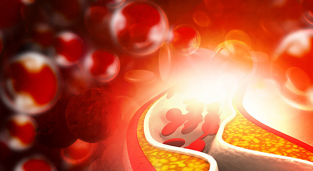
Peripheral arterial disease (PAD), also known as peripheral vascular disease (PVD), is a very common condition that affects 10 million Americans, and 12-20 percent of Americans age 65 and older, each year. PAD develops most commonly as a result of atherosclerosis (hardening of arteries in the heart). It occurs when cholesterol and scar tissue build up inside the arteries, forming a substance called plaque.
As plaque narrows and clogs the arteries, it can create a very serious condition that decreases blood flow to the legs, which can result in pain when walking, and can lead to gangrene and possible amputation.
People with PAD are at increased risk for heart disease, aortic aneurysm and stroke. PAD is also a marker for diabetes, hypertension and other conditions.
Detection and diagnosis
Some signs of narrowed, enlarged or hardened arteries may be found during a physical exam. These include:
- A weak or absent pulse below the narrowed area of the artery
- Decreased blood pressure in an affected limb
- Whooshing sounds (bruits) over the arteries, heard with a stethoscope
- Signs of a pulsating bulge (aneurysm) in the abdomen or behind the knee
- Evidence of poor wound healing in the area where blood flow is restricted
Depending on the results of a physical exam, your doctor may suggest one or more diagnostic tests, including:
Doppler ultrasound
This test involves the use of a special ultrasound device to measure blood pressure at various points along the arm or leg. These measurements can help the physician gauge the degree of any blockages, as well as the speed of blood flow in your arteries.
Ankle-brachial index
Comparing the blood pressure in the ankle with the blood pressure in the arm is known as the ankle-brachial index. An abnormal difference may indicate peripheral vascular disease, which is usually caused by atherosclerosis.
Electrocardiogram
An electrocardiogram (ECG) records electrical signals as they travel through your heart. An ECG can often reveal evidence of a previous heart attack or one that’s in progress. If signs and symptoms occur most often during exercise, the physician may ask you to walk on a treadmill or ride a stationary bike during an ECG.
Arteriogram
To better view blood flow through the heart, the patient’s artery is injected with a special dye under X-ray. This is known as an arteriogram. The images outline narrow spots and blockages, which can also be repaired or treated during the same procedure.
Other imaging tests for cardiovascular care
The use of other imaging techniques such as a CT scan and MRI to evaluate the arteries is becoming more common. They are less invasive than the conventional arteriogram because there is no need for arterial puncture or sedation. The studies are called CTA (CT Arteriogram) or MRA (MR Arteriogram), and images can be provided in 3D reconstruction for evaluation of hardening and narrowing of large arteries, as well as aneurysms and calcium deposits in the artery walls.
Treatment options
After PAD has been diagnosed, several treatment options are available, including:
- Medication
- Endarterectomy
- Bypass surgery
- Interventional cardiology
- Thrombolytic therapy
- Angioplasty and stenting
- Diamondback rotablator

