Noninvasive Diagnostic Procedures
At Valley Hospital Medical Center, a wide range of advanced tests and procedures are available to help diagnosis and detect a variety of cardiovascular conditions and disorders. These tests can provide doctors with information about heart valves, the heart muscle, coronary arteries and more.
Echocardiography
Echocardiography (also known as an echocardiogram) is a type of ultrasound diagnostic tool that uses high-pitched sound waves to produce an image of the heart. Sound waves are sent through a transducer that is placed on the patient’s chest. Sound waves reflect off certain areas of the heart and are then converted into pictures. This test is used to check the heart's ability to pump blood effectively throughout the body and measure how much blood is actually pumped in each contraction. It can also be used to evaluate the valve function, find evidence of heart failure and confirm that the heart is of normal size.
There are three main types of echocardiograms:
Transesophageal (TEE)
This is a specialized test that emits sound waves through the esophagus instead of the chest or abdomen. This gives the physicians better pictures of the heart because the transducer is closer to the heart. Local and intravenous anesthetics are used with TEE to minimize any discomfort to the back of the throat.
Stress Echocardiogram
In this test, patients exercise on a treadmill, or are given a drug to place stress on the heart. An echocardiogram is performed before and after the heart is overloaded in order to see if the patient has a significantly reduced blood flow to the heart.
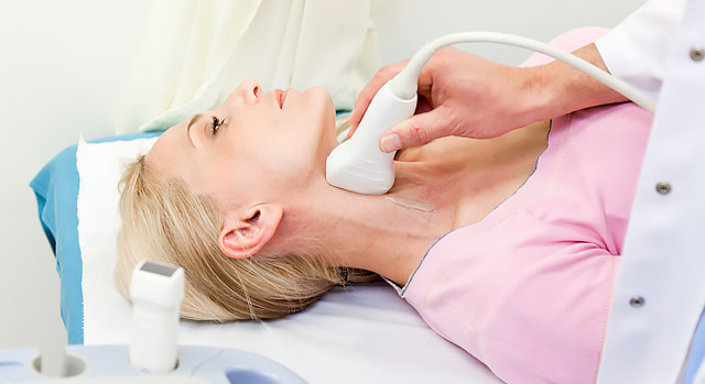
Carotid Ultrasound
This is a test that looks at plaque build-up in the arteries of the neck supplying blood to the brain to give physicians information about risk of stroke, and to predict higher likelihood of plaque in other artery branches including the heart, kidneys and legs.
Carotid ultrasounds compare pressure in the arms and legs and can help identify peripheral vascular disease and increased risk of cardiovascular events, including heart attack and stroke.
Radionuclide Imaging
Radionuclide imaging is a technique in which small amounts of radioactive materials are injected into a patient’s body to help physicians obtain detailed images that can show parts of the heart muscle with abnormal blood flow. These images can be particularly helpful in discovering blocked heart arteries and damaged heart muscle.
Cardiac MRI
Magnetic resonance imaging (MRI) provides detailed pictures without the need for any radiation or injected dyes. By using a powerful magnetic field and radio signals, an MRI provides detailed images of blood vessels and how blood is flowing throughout the body.
Chest X-ray
This test provides images of the heart, the blood vessels in the chest and the diaphragm. Chest X-rays can provide images from many different angles of the chest, and are used to help diagnose and examine conditions of heart disease.

